

Retinal Detachment Surgery Cost in India
Last updated: May 3, 2026
Starting from USD 2,200
The Retinal Detachment Surgery Cost in India starts from USD 2,200. It varies depending on the type of treatment, the patient’s medical condition, the doctor, the facility, and the city where you choose to get the surgery done.
The cost quoted above is indicative and should not be taken as the final cost of the surgery. The final cost of retinal detachment surgery in India can be ascertained after the surgeon in India has physically evaluated the patient. The cost in Indian Rupees can vary based on the exchange rate.
INCLUDES
- Surgery
- Stay at the Hospital
- Pre-operative Investigations
- Medicines, Consumables
- Food at the hospital
- Airport Transfers
- MediTraWell assistance
DOES NOT INCLUDE
- Accommodation outside the Hospital
- Air tickets
- Medical Visa fee
STAY REQUIRED
- Stay at the Hospital - 1 to 2 days
- Stay at the Hotel - 7 to 8 days
- Stay in India - 8 to 10 days
Factors that affect Retinal Detachment Surgery Cost in India:
- Type of Treatment Required:
While some patients may do well with vitrectomy or cryopexy to treat retinal detachment, others may need pneumatic retinopexy to treat the condition. Our expert doctors will advise the right treatment for you depending on the severity of your condition, which in turn will affect the cost of retinal detachment surgery in India.
- Ophthalmologist’s Fees
A significant component of the retinal detachment surgery cost in India is the ophthalmologist’s fees. IndiCure recommends highly experienced, skilled, board-certified surgeons who have the track record of delivering excellent results. While the charges may vary based on the surgeon’s experience, you can be confident that you are in safe and capable hands when opting for eye surgery in India with IndiCure Health Tours.
- Your Choice of Surgical Facility
Selecting an accredited medical facility with skilled and qualified medical staff is essential for the success of your retinal detachment surgery in India. Larger cities in India generally provide superior medical facilities and more experienced surgeons, leading to relatively higher costs as compared to some smaller cities in India. IndiCure Health Tours recommends surgical facilities in these larger cities to prioritize quality of care and ensure safety of our international patient guests.
- Treatment-Related Expenses
The surgery-related expenses include the pre- and post-surgical expenses. The pre-surgical expenses are associated with the age and medical condition of the patient and thus the number and type of investigations required. Post-surgical expenses may include prescription medications and follow-up consultations.
We at MediTraWell, understand that you travel with a budget in mind and do not like to be greeted by surprises after arrival in India. We thus club all these expenses and give you a package cost that is inclusive and affordable at the same time.
Your case manager shall give you an estimated cost of your surgery after discussing your medical reports with the surgeon. The final cost, however, shall be confirmed after your consultation with the surgeon.
Medi Trawell Services
Making Medical Travel to India Affordable & Hassle-free for 15+ Years

We Help you Choose the Right Treatment, Surgeon & Hospital

We Arrange Video/Telephonic Consultation with the Surgeon

We Assist you with Visa & Accommodation

We Receive you at the Airport and Drop you at Hotel/Hospital

We Assist you the at Hospital & Provide Post Operative Support
Our services are FREE for our patients.
In fact, we have Special Negotiated Rates with the Hospitals and you can avail Discounted Rates when you choose to Travel with MediTrawell. Container
Best Ophthalmologists in India

Dr. Nishikant Borse
Retinal Surgery
MBBS, MS, FMRF, FASRS
Mumbai
Dr. Nishikant Borse is not only regarded as one of the best ophthalmologists in Mumbai, but also one of the best eye doctors in India. With more than 25 years of experience and more than 10,000 surgeries to his credit with challenging retina cases, he is one of India’s top retina surgeons. After completing his medical studies at the illustrious Grant Medical College in Mumbai and earning a Master’s degree in Ophthalmology, he underwent advanced training and Fellowship at the prestigious Sankara Nethralaya in Chennai.
Dr. Borse is frequently asked to speak as a guest speaker at conferences all around the country and the world. He has led various training sessions for teaching the complexities of retinal surgery in Mumbai and other areas of the country. He has carried out real retinal procedures at numerous conferences. He has had a number of research papers published in both domestic and foreign journals.
Dr. Borse has been many awards nationally and internationally, like The Rhett Buckler Award by the American Society of Retina Specialists, Cesare Forlini Award by the European Vitreoretinal Society, C S Reshmi Award by the All India Ophthalmological Society,
He is a member of the Bombay Ophthalmologists Association, the Maharashtra Ophthalmological Society, the All India Ophthalmological Society, the Vitreo-Retinal Society of India, the American Academy of Ophthalmology, and the International Society of Ocular Trauma. He is also a Fellow of the American Society of Retina Specialists.
American Academy of Ophthalmology, and the International Society of Ocular Trauma. He is also a Fellow of the American Society of Retina Specialists
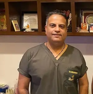
Dr Suraj Munjal
LASIK
MBBS, MS
Delhi
Dr Suraj Munjal is a well renowned name in Eye care not only in Delhi NCR, but the entire North India. With proficiency in refractive surgeries and corneal transplants, Dr Munjal is considered one of the best eye doctors in India and manages a wide array of complex and referred cases from across India and other countries. He is widely regarded as the best eye doctor in Delhi and has amassed extensive experience in performing successful LASIK surgeries.
With his dedication to delivering exceptional eye care, Dr Munjal is committed to providing affordable and personalized treatments without compromising on ethics. His commitment has led him to perform surgeries in various Middle Eastern countries, including Iraq, Dubai, Kurdistan, and Bahrain, at the invitation of local governments.
Best Eye Hospitals in India
Nova IVF Infertility, New Delhi
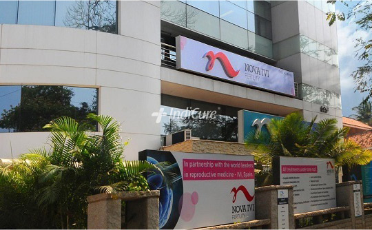
Location: New Delhi
Speciality: Infertility & IVF
Loading...
Loading...
Southend Fertility and IVF is now Nova IVF Fertility. It offers a complete Infertility Management program to couples who are experiencing difficulty having a child. The facility has been a centre of expertise for couples seeking fertility therapy in Delhi for over 20 years. The clinic has also taught thousands of gynecologists who now work as fertility specialists. The clinic claims complete treatment openness, sympathetic staff, a vibrant environment, and hopeful outcomes. Patients who return to the clinic after failed cycles demonstrate their trust in and satisfaction with their treatment method.
Apollo Cradle Royale, Nehru Place, Delhi

Location: New Delhi
Speciality: Women & child care, IVF
Accreditation: Accreditation : NABH
No. of Beds: 18
Apollo Cradle Royale in Delhi excels in Gynaecology, Maternity, Paediatrics, Paediatric Surgery, and Neonatology, serving as a centre of excellence in these specialized healthcare areas. Spanning an expansive 35,000 square feet, Apollo Cradle is a premium healthcare facility that provides a wide range of services. The hospital offers procedures such as IVF, IUI, fertilization, IVF-ET, IUD placement, laparoscopic sterilization, complex pregnancy treatment, gynecological endoscopy/laparoscopy, abdominal hysterectomy, clitoral hood reduction, and hysterectomy (both abdominal and vaginal). Apollo Cradle boasts an advanced level III Neonatal Intensive Care Unit (NICU) that offers top-notch care. Additionally, for women requiring emergency care, there is an additional Intensive Care Unit (ICU).
Questions you must ask
Here is a set of questions you should consider asking before commencing your medical journey for retinal detachment surgery in India.
- Is the eye surgeon board certified?
- How experienced is the Surgeon?
- Which language does the surgeon speak?
- Is the surgery done in a well-equipped facility?
- Can you give me any information on outcomes and complication rates?
- How much pain can I expect, and how will it be managed in the hospital and after I go home?
- What surgical option is recommended for me?
- What about the risks involved?
- Does the surgeon use a certified anesthetist?
- How long will the recovery period be?
Be ready to respond about:
Be ready to respond about:
- Medical history and exams
- Previous surgeries
- Current medication review
- History of smoking, drugs, or alcohol
Retinal Detachment
A retinal detachment is a medical emergency in which a thin layer of tissue at the back of the eye (the retina) slips away from its normal location.
The retinal cells are separated from the layer of blood vessels that give oxygen and sustenance by retinal detachment. The longer you wait to treat a retinal detachment, the more likely you are to lose vision in the affected eye permanently.
The abrupt emergence of floaters and flashes, as well as impaired vision, are all possible symptoms of retinal detachment. Contacting an ophthalmologist (eye doctor) as soon as possible will help you save your vision.
Symptoms of Retinal Detachment:
Retinal detachment is painless. However, there are virtually always warning signals before it develops or has progressed, such as:
- The unexpected emergence of a large number of floaters – little specks that appear to wander across your field of vision
- Light flashes in one or both eyes (photopsia)
- Hazy vision
- Reduced side (peripheral) vision with time
- Curtain-like shadow over your visual field
Types of Retinal Detachments:
Rhegmatogenous: These are the most common types of retinal detachments. A hole or tear in the retina allows fluid to travel through and gather behind the retina, forcing the retina away from the underlying tissues, causing rhegmatogenous detachments. The portions of the retina that detach lose blood supply and stop operating, resulting in visual loss.
Rhegmatogenous detachment is most commonly caused by aging. The vitreous, a gel-like fluid that fills the inside of your eye, may alter in consistency and shrink or become more liquid as you become older. The vitreous normally separates from the retina’s surface without causing any problems, a situation known as posterior vitreous detachment (PVD). A tear is one of the complications of this separation.
The vitreous may pull on the retina with enough force to cause a retinal tear as it separates or peels away from the retina. If the tear is not repaired, the liquid vitreous can leak into the space behind the retina, causing the retina to detach.
Tractional: When scar tissue forms on the surface of the retina, the retina pulls away from the back of the eye, causing detachment. People with poorly controlled diabetes or other diseases are more likely to develop tractional separation.
Exudative: Fluid collects behind the retina in this type of detachment, but there are no holes or tears in the retina. Age-related macular degeneration, eye injuries, malignancies, and inflammatory illnesses can all produce exudative detachment.
Risk Factors for Retinal Detachment
The following factors raise your chances of developing a retinal detachment:
- Aging- Retinal detachment is more likely in adults over 50 years old.
- Previous retinal detachment in one eye
- Retinal detachment in the family
- Nearsightedness to the extreme (myopia)
- Previous eye surgery, such as cataract removal
- Previous eye damage that was severe, such as Retinoschisis, uveitis, or weakening of the peripheral retina
Treatment for Retinal Detachment
A retinal tear, perforation, or detachment is usually invariably repaired with surgery. There are a variety of strategies to choose from. Inquire about the risks and advantages of your treatment options with your ophthalmologist.
Retinal Tears
Your eye surgeon may recommend one of the following procedures to prevent retinal detachment and maintain vision if a retinal tear or hole hasn’t proceeded to separation.
Laser surgery
procedure that involves the use of (photocoagulation). A laser beam is sent into the eye through the pupil by the surgeon. The laser creates scarring around the retinal tear, which “welds” the retina to the underlying tissue.
Freezing (cryopexy)
After numbing your eye with a local anesthetic, the surgeon uses a freezing probe to freeze the outer surface of your eye exactly above the tear. The freezing creates a scar that aids in the retina’s attachment to the eye wall.
You will be advised to avoid activities that may affect the eyes, such as running for a couple of weeks.
Retinal Detachment
If your retina has detached, you’ll require surgery to restore it as soon as possible after receiving your diagnosis. The type of surgery recommended by your surgeon will be determined by a number of criteria, including the severity of the separation.
Air or gas is injected into your eye-The surgeon injects a bubble of air or gas into the center of the eye in a procedure known as pneumatic retinopexy. The bubble, when appropriately positioned, presses the portion of the retina containing the hole or holes against the eye’s wall, preventing fluid from flowing into the space behind the retina. During the operation, your doctor may employ cryopexy to heal the retinal break.
Fluid that has accumulated beneath the retina is absorbed by itself, allowing the retina to cling to the eye’s wall. To keep the bubble in the appropriate position, you may need to hold your head in a specific position for several days. The bubble will eventually dissolve on its own.
Indenting the surface of your eye– The surface of your eye is indented. The surgeon sews (sutures) a piece of silicone material to the white of your eye (sclera) over the damaged area in a process known as scleral buckling. This treatment indents the eye’s wall, relieving part of the tension exerted on the retina by the vitreous.
Your surgeon may make a scleral buckle that encircles your entire eye like a belt if you have multiple tears or holes or an extensive separation. The buckle is positioned in such a way that it does not obstruct your vision, and it usually stays in place indefinitely.
Draining and replacing the fluid in the eye– The fluid in the eye is drained and replaced. Vitrectomy is the operation in which the surgeon removes the vitreous as well as any tissue that is tugging on the retina. The vitreous area is then injected with air, gas, or silicone oil to assist flatten the retina.
The air, gas, or liquid will eventually be absorbed, and the vitreous area will be replenished with bodily fluid. Silicone oil may be surgically removed months later if it is used.
A scleral buckling treatment can be done with a vitrectomy.
It may take several months for your vision to improve after surgery. For a successful treatment, you may require a second surgery.
When is the Treatment for Retinal Detachment Needed?
If you are suffering the signs or symptoms of retinal detachment, seek medical help right away. Retinal detachment is a medical emergency that can result in lifelong eyesight loss.
Surgery Information
| Surgery Time | 1-2 hours |
|---|---|
| Anesthesia Type | IV Sedation |
| Hospitalization | 1-2 days |
| Stitch Removal | 3 weeks |
| Recovery Time | 4-6 weeks |
Surgical Procedure
Vitrectomy
Step 1
Three small ports are created in the white (sclera) of your eye. One allows a continuous flow of fluid into your eye, called an ‘infusion.’ The second port is for a fiber-optic ‘light pipe’ to illuminate your eye. The third port is for tools needed during surgery, like a ‘cutter’ to remove the vitreous.
Step 2
After that, cryotherapy will be used. During the surgery, the fluid inside your eye (vitreous humor) is replaced with air. When the air is initially injected, you might hear a whistling sound. At the end of the operation, this air will be replaced with a gas, which is commonly C3F8 or SF6.
Step 3
To protect your eye and aid in the healing process, a pad, and a rigid shield will be placed over your eye. The pad provides cushioning and absorbs any fluids, while the shield acts as a barrier against accidental bumps or pressure, ensuring your eye remains undisturbed.
What Results can I expect from Retinal detachment surgery?
Scleral buckling, pneumatic retinopexy, and primary vitrectomies with or without buckles have all been shown to be helpful in healing retinal detachments.
With detachment surgery today, our doctors expect a primary success rate of 90% or higher and a final reattachment rate of over 95%.
What is the Recovery after Retinal detachment surgery like?
The vision may take several weeks or months to fully recover, but the majority of the discomfort will go away within the first week. Before resuming your typical activities, you’ll need 2 to 4 weeks to heal. However, everyone recovers at their own speed. To improve as rapidly as possible, you should follow the instructions given by your doctor.
You will be given antibiotic and anti-inflammatory drops to take for a few weeks after ICL surgery. Most patients can return to normal activities within a week of ICL surgery, but you should not drive until you have been told it is safe.